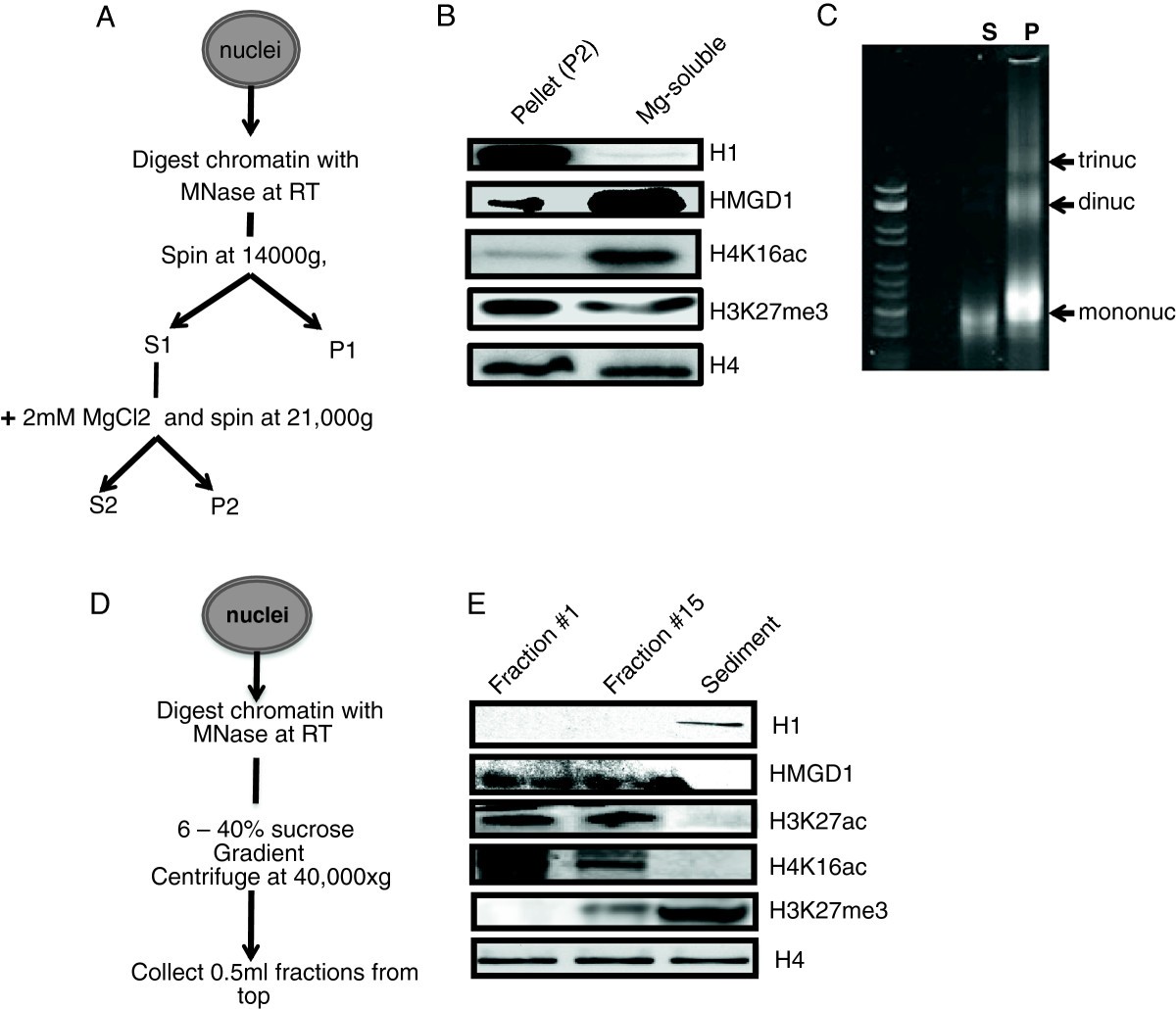
Figure 1

HMGD and H1 are associated with euchromatic and heterochromatic fractions respectively. (A) Salt fractionation of chromatin procedure. (B) Western blot of H1 and HMGD1 in heterochromatic (P2) and euchromatic (S2) chromatin fractions. We digested chromatin from S2 nuclei with micrococcal nuclease and fractionated it to yield supernatant (S2) and pellet (P2) fractions. The western blot shows that H1 is primarily found in the heterochromatic fraction (P2) and HMGD1 is primarily found in the euchromatic fraction (S2). The histone mark H4K16ac is a positive marker for active euchromatin and the histone H4 antibody is used as a chromatin loading marker. (C) Ethidium bromide stained 3.3% Nusieve™ agarose gel showing the DNA associated with the S2 (magnesium-soluble) and P (magnesium-insoluble) fractions. (D) Sucrose gradient fractionation procedure. (E) Western blot analysis of H1 and HMGD1 released from MNase-digested chromatin from S2 cells. The 6 – 40% sucrose gradient reveals that H1 is bound to the heavier heterochromatin, while HMGD1 is bound to the lighter euchromatin.